Facilities and Equipment
Equipment
According to the facilities and needs for each laboratory, the following equipment are available in the pathology laboratory at the Pathology Department and the Pathology laboratories in Mansoura University Centers and Hospitals:
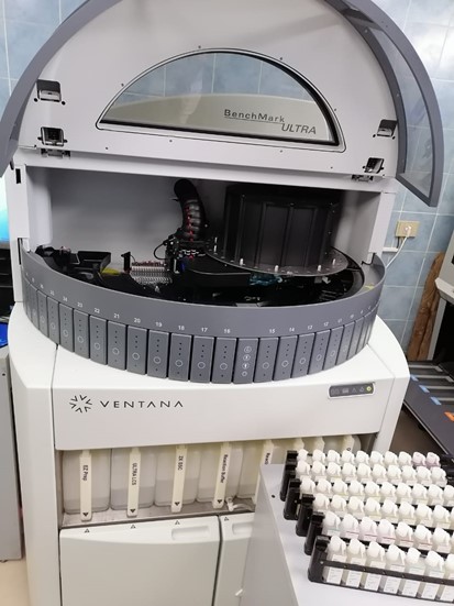
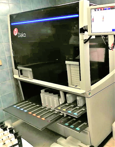
- Automated and semi-automated autostainers for immunohistochemical staining.
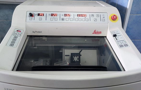
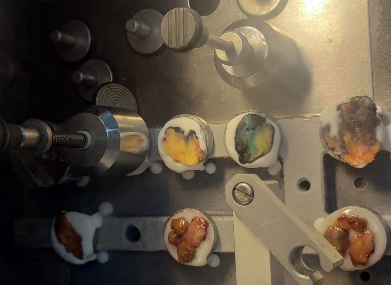
- Cryostats for frozen section preparation.
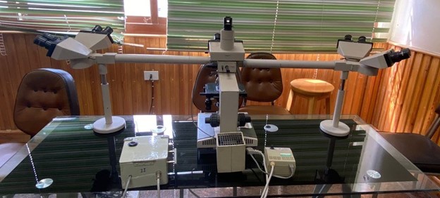
- Binocular, and multi-head ordinary-light microscopes for slide examination.
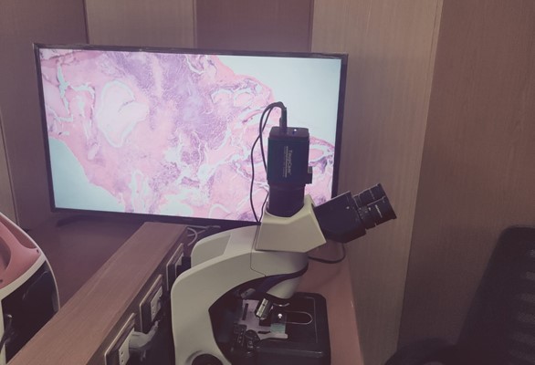
- Microscope systems-equipped with high-resolution digital cameras to show and photograph slides.
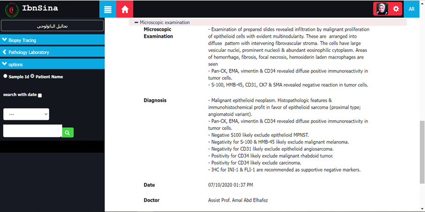
- Computers and printers connected to the Internet with access to the "Ibn Sina" medical records system to export pathology reports as soon as they are written, and confirmed, and to store data directly in the system's databases.
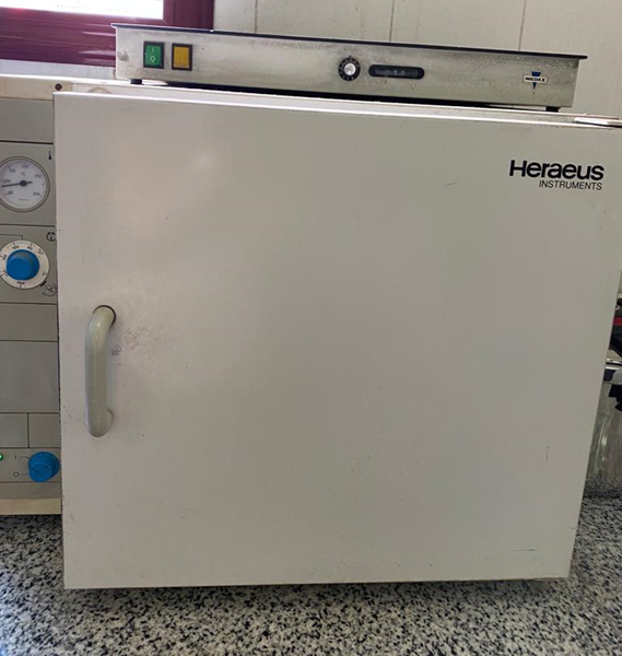
- Ovens and microwaves.
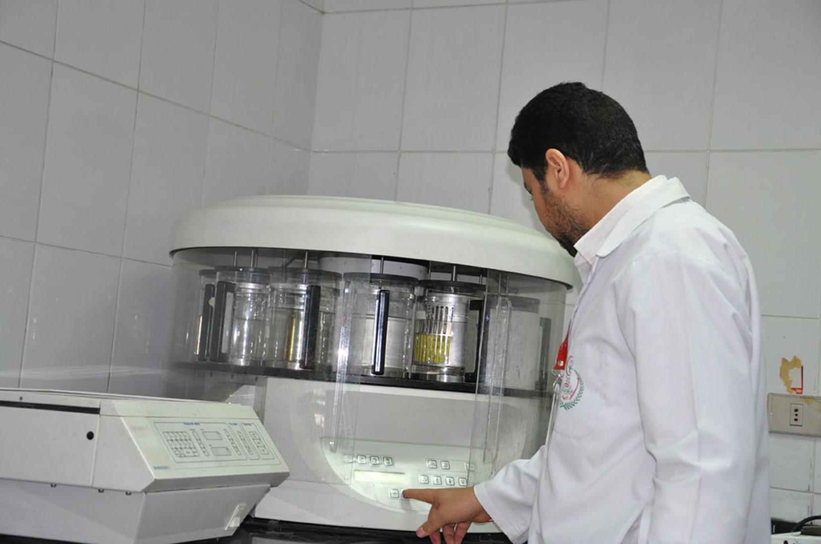
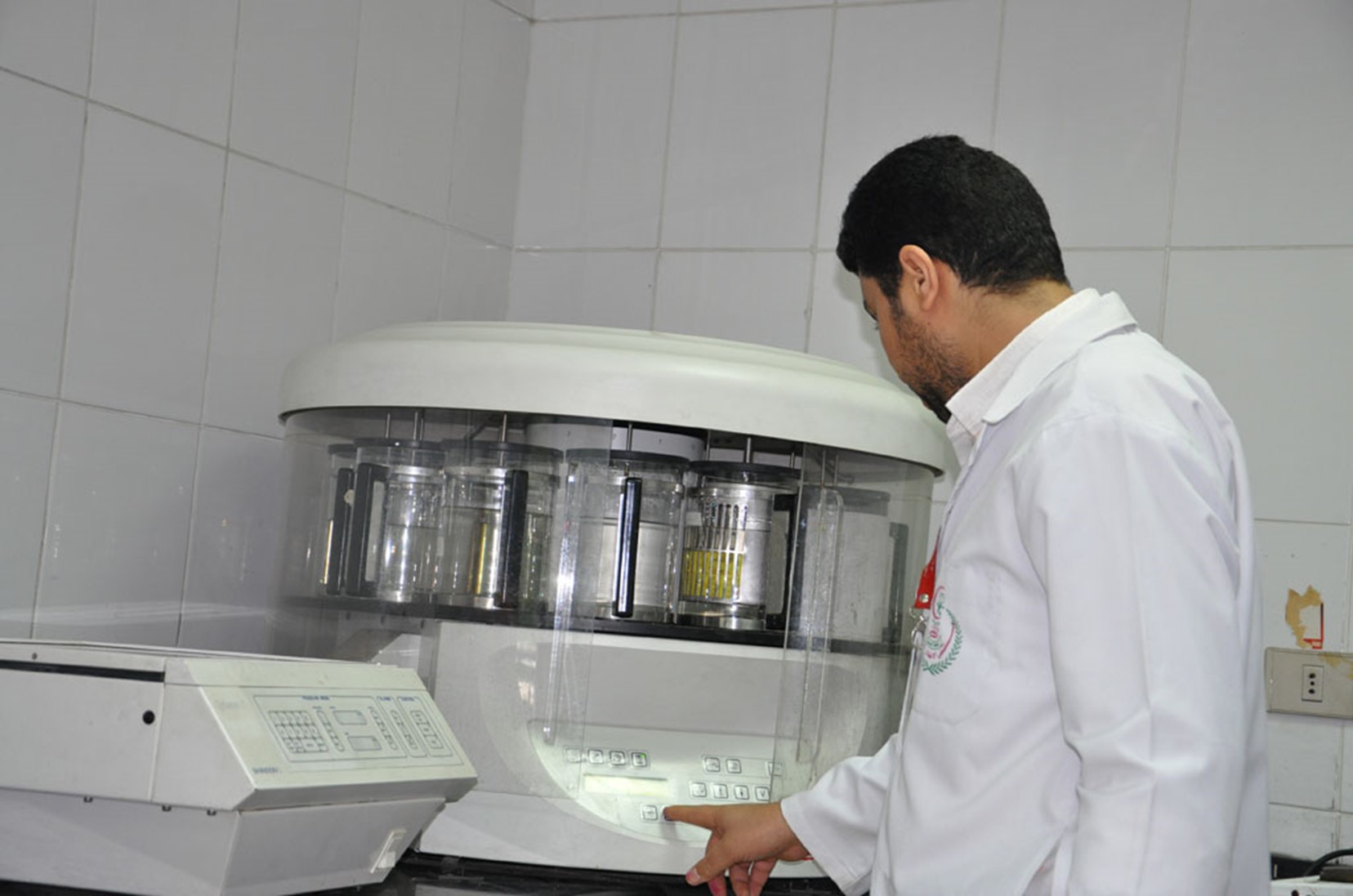
- Tissue processors to prepare the dissected tissue samples using the paraffin technique.
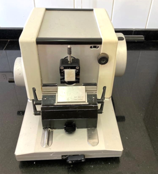
- Microtomes for preparing tissue sections.
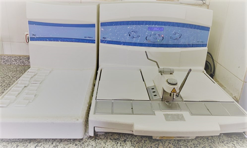
- Paraffin-embedding and cooling apparatus.
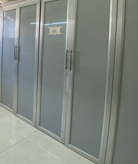
- Refrigerators for chemicals and immunohistochemical antibodies and kits.
- Centrifuge for fluid samples.
- Storage cabinets for keeping surgical specimens after dissection.
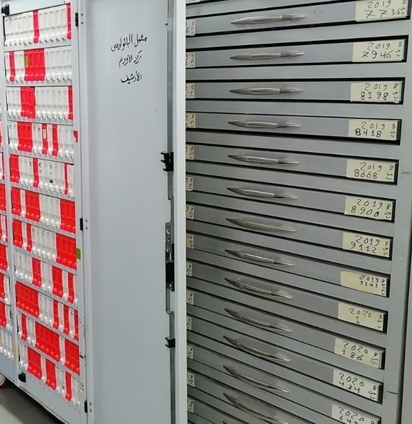
- Storage cabinets designed for archiving microscopic glass slides and paraffin tissue-blocks.
Available Pathological Examinations
In the pathology laboratory at the Pathology Department and the pathology laboratories in Mansoura University Centers and Hospitals, we have the ability to perform the following examinations according to the lab specializations, equipment and facilities:
- Microscopic examination of surgical specimens including core, incision and excision biopsies using the routine hematoxylin and eosin staining on paraffin sections.
- Performing various types of diagnostic and prognostic immunohistochemical stains, especially that governs therapeutic approaches, such as hormone receptors, epidermal growth factor receptors, and Ki-67 proliferation index in cases of breast cancer.
- Cytological examination for different fluid samples with the possibility of preparing paraffin cell-blocks and applying immunocytochemistry on it.
- Frozen-section preparation and examination during surgery for provisional intraoperative pathological diagnosis.
- Examination and immunophenotyping of bone marrow core biopsies in different hemato-lymphoid disorders and comparing the biopsy findings to the flow-cytometry for appropriate diagnosis.
- Examination of renal biopsies and applying special stains to reach the most accurate diagnosis.
- Examination of liver biopsies in the different pathological conditions, including biopsies from both the donor and the recipient in cases of liver transplantation, and the follow-up liver biopsies to assess transplant rejection as well.
Facilities
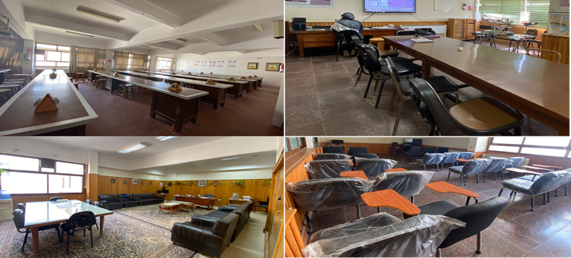
- Student laboratories.
- Sample preparation laboratories.
- Dissection rooms.
- Department council room.
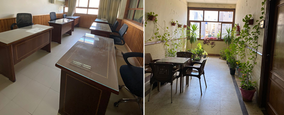
- Staff offices and assistant staff rooms.
- Seminar and post-graduate lecturing hall.
- Microscopic examination room.
- Departmental secretarial room.
- Secretarial room for specimen reception and registration, and writing reports.
- Break area.
